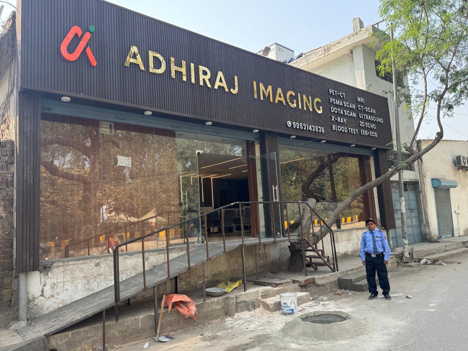
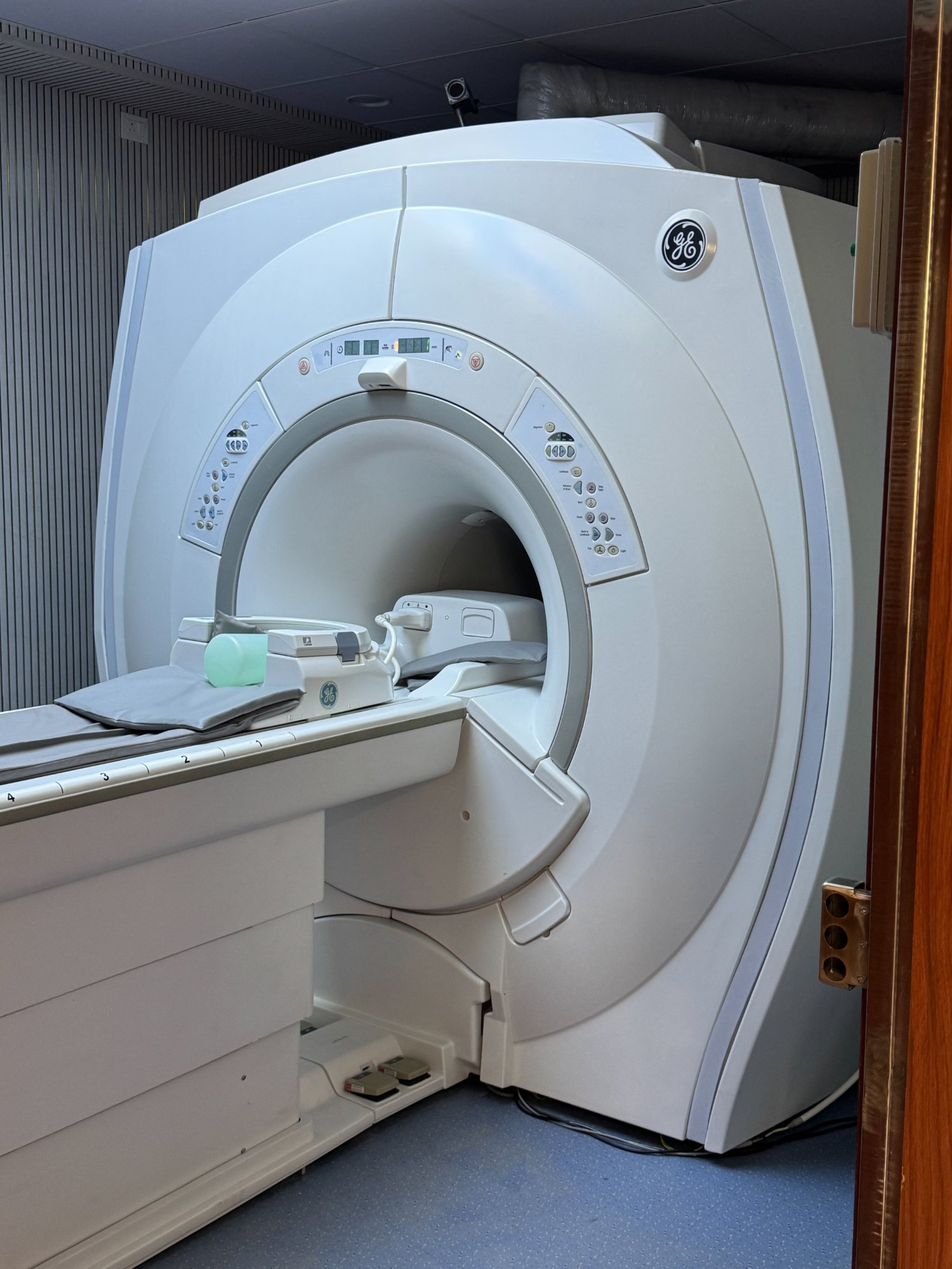
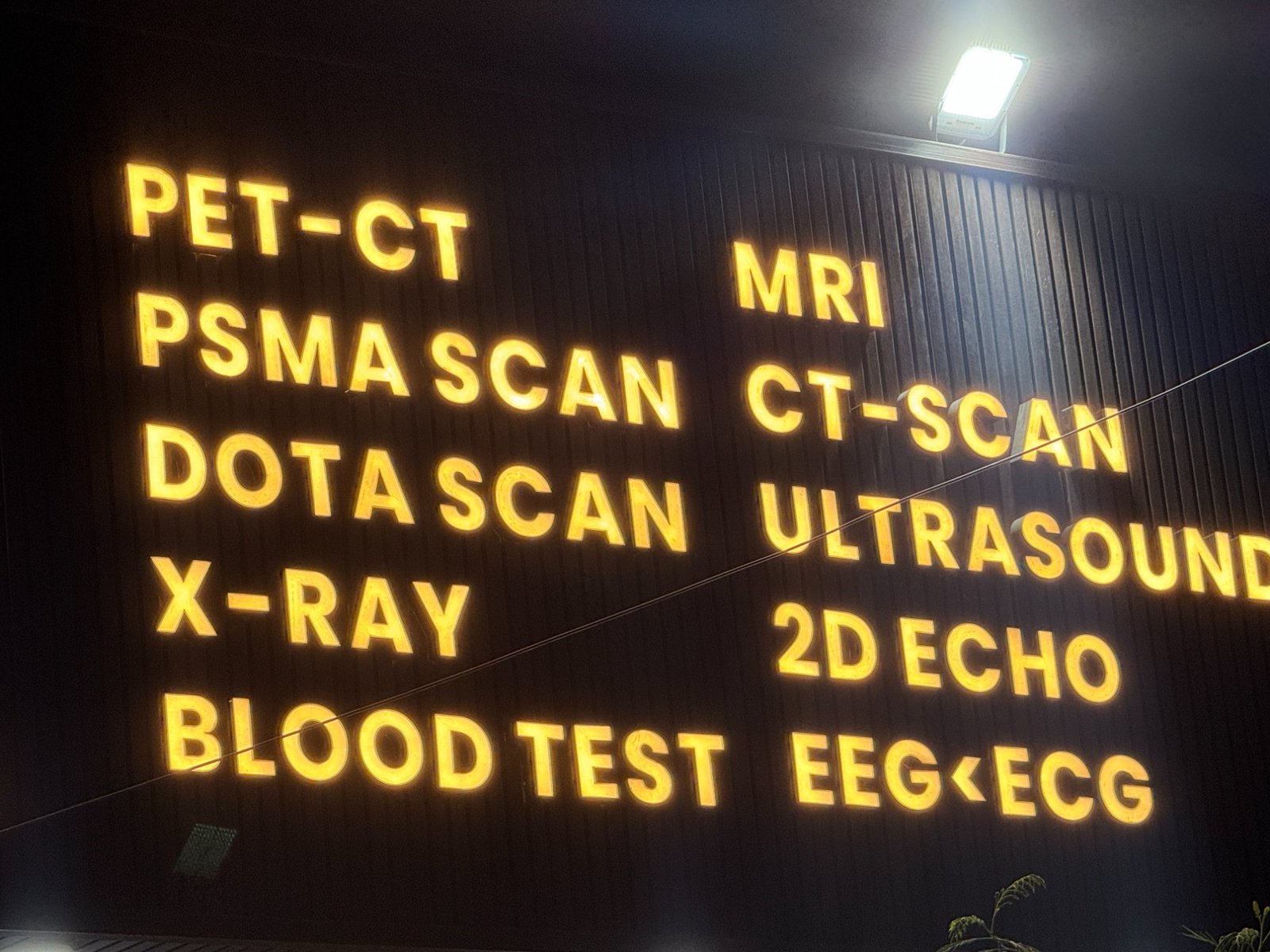
ADHIRAJ IMAGING, DILSHAD GARDEN
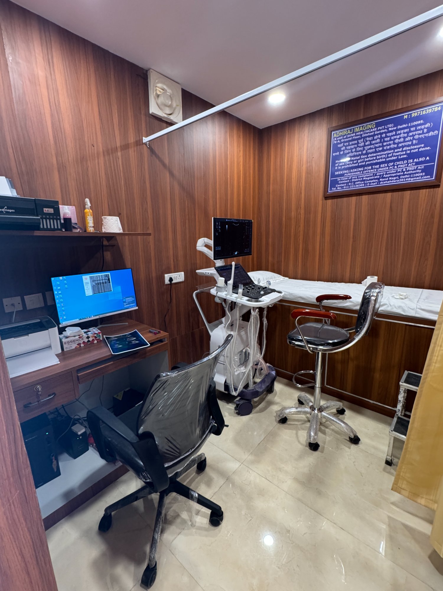
ECHO ROOM

WAITING AREA
GE LOGIQ SYSTEM
RECEPTION
What is a 2D Echo?
A Live Ultrasound of Your Beating Heart
A 2D Echo (Two-Dimensional Echocardiography) is an ultrasound scan of the heart. Using high-frequency sound waves, it produces real-time moving images of the heart's chambers, valves, walls and major blood vessels — giving your cardiologist a live picture of how your heart is functioning.
Unlike an ECG, which records only the heart's electrical activity, a 2D Echo shows the heart's actual physical structure and mechanical movement — how strongly the heart muscle contracts, whether valves open and close correctly, and whether any fluid surrounds the heart.
It is completely painless, non-invasive, uses no radiation, and requires no preparation. The test takes 20–30 minutes and reports are delivered the same day via WhatsApp.
ECG vs 2D Echo — Key Difference
ECGRecords electrical activity only — rhythm, rate, conduction
2D EchoShows heart structure, movement, valves and blood flow in real time
Colour DopplerMaps blood flow direction and speed through each valve
RadiationNone — completely safe ultrasound technology
Book 2D Echo / Echocardiography
or book instantly
WhatsApp Us
📍 76-A, GF1, Opp. IHBAS Hospital Gate No. 3, Dilshad Garden, Delhi – 110095
⏰ Mon–Sun · 8:00 AM – 8:00 PM
🚇 5 min from Dilshad Garden Metro (Red Line)
⏰ Mon–Sun · 8:00 AM – 8:00 PM
🚇 5 min from Dilshad Garden Metro (Red Line)